
OP64
Anti-p21WAF1 (Ab-1) Mouse mAb (EA10)
liquid, clone EA10, Calbiochem®
别名:
Anti-CIP1, Anti-SD11, Anti-p21, Anti-WAF
登录 查看组织和合同定价。
选择尺寸
变更视图
关于此项目
NACRES:
NA.43
UNSPSC Code:
12352203
Clone:
EA10, monoclonal
Species reactivity:
human
Application:
—
Citations:
59
biological source
mouse
Quality Level
antibody form
purified antibody
antibody product type
primary antibodies
clone
EA10, monoclonal
form
liquid
contains
≤0.1% sodium azide as preservative
species reactivity
human
should not react with
mouse, rat
manufacturer/tradename
Calbiochem®
storage condition
do not freeze
dilution
(Flow Cytometry (2 µg/mL)
Frozen Sections (5 µg/mL)
Immunoblotting (1-3 µg/mL)
Immunofluorescence (1-5 µg/mL)
Immunoprecipitation (2 µg/sample)
Paraffin Sections (5 µg/mL or use OP64F; heat pre-treatment required; and application references))
isotype
IgG1
shipped in
wet ice
storage temp.
2-8°C
target post-translational modification
unmodified
Gene Information
human ... CDKN1A(1026)
General description
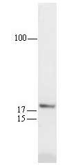
Recognizes the ~21 kDa p21WAF1 protein in skin and colon tissue and in cells expressing wild-type p53 (e.g. Hs27 or U205 cells treated with DNA damaging agents).
This Anti-p21WAF1 (Ab-1) Mouse mAb (EA10) is validated for use in FC, Immunoblotting, IF, IP, Paraffin Sections for the detection of p21WAF1 (Ab-1).
Application
Flow Cytometry (2 g/ml or use Cat. No. OP64F; see application references)
Frozen Sections (5 g/ml or use Cat. No. OP64F)
Immunoblotting (1-3 g/ml)
Immunofluorescence (1-5 g/ml or use Cat. No. OP64F)
Immunoprecipitation (2 g/sample)
Paraffin Sections (5 g/ml or use OP64F; heat pre-treatment required; see comments and application references)
Packaging
Please refer to vial label for lot-specific concentration.
Analysis Note
Positive Control
Any cell line expressing wild-type p53 (e.g. Hs27 or U2OS treated with DNA-damaging agents) or skin or colon tissue
Any cell line expressing wild-type p53 (e.g. Hs27 or U2OS treated with DNA-damaging agents) or skin or colon tissue
Other Notes
Agarwal, M.L., et al. 1995. Proc. Natl. Acad. Sci. USA92, 8493.
Chen, Y.Q., et al. 1995. Int. J. Oncology7, 889.
Deng, C., et al. 1995. Cell82, 675.
El-Deiry, W.S., et al. 1995. Cancer Res.55, 2910.
Waldman, T., et al. 1995. Cancer Res.55, 5187.
Elbendary, A., et al.1994. Cell Growth Diff.5, 1301.
El-Deiry, W.S., et al. 1994 Cancer Res.54, 1169.
Li, R., et al. 1994. Nature371, 534.
Michieli, P., et al. 1994. Cancer Res.54, 3391.
Noda, A., et al.1994. Exp. Cell Res.211, 90.
El-Deiry, W.S., et al.1993. Cell75, 817.
Gu, Y., et al. 1993. Nature366, 707.
Harper, J.W., et al.1993. Cell75, 805.
Xiong, Y., et al.1993. Genes Devel.7, 1572.
Xiong, Y., et al.1993. Nature366, 701.
Xiong, Y., et al.1992. Cell71, 505.
Maximal p21WAF1 expression requires wild type p53 activity. Treatment of U2OS or MCF7 cells with DNA damaging agents (such as doxorubicin at 0.2 µg/ml) induces wild type p53 expression which in turn activates WAF1 expression. Serum stimulation of quiescent cells will give low level WAF1 expression independent of p53 expression. Untreated cells will express little p21WAF1 and can be used as a negative control. Cat. No. OP64F was tested in HALT cells induced by incubation at 31°C; FITC-goat anti-mouse IgG (Cat. No. DC13L) was used as a negative control. This antibody will immunoprecipitate p21WAF1 but not associated proteins. For immunoblotting applications, use a 0.22 µm filter and visualize by chemiluminescence. For staining paraffin sections, heating the tissue in 10 mM citrate buffer is required (see application references). In either paraffin or frozen sections of normal human colon, the non-dividing cells of colonic epithelium will stain positive for p21WAF1 while the proliferating compartment of crypts will not stain. Antibody should be titrated for optimal results in individual systems.
Legal Information
CALBIOCHEM is a registered trademark of Merck KGaA, Darmstadt, Germany
Disclaimer
Toxicity: Standard Handling (A)
Still not finding the right product?
试用我们的 产品选型工具 工具缩小选择范围
存储类别
11 - Combustible Solids
wgk
WGK 1
flash_point_f
Not applicable
flash_point_c
Not applicable
null
Elevated MDM2 boosts the apoptotic activity of p53-MDM2 binding inhibitors by facilitating MDMX degradation.
Xia, et al.
Cell Cycle, 7, 1604-1612 (2020)
Vladimir A Shamanin et al.
Molecular and cellular biology, 24(5), 2144-2152 (2004-02-18)
Inactivation of the ARF-p53 tumor suppressor pathway leads to immortalization of murine fibroblasts. The role of this pathway in immortalization of human epithelial cells is not clear. We analyzed the functionality of the p14(ARF)-p53 pathway in human mammary epithelial cells
全球贸易项目编号
| 货号 | GTIN |
|---|---|
| OP64-20UG | 04055977227291 |
| OP64-100UG | 07790788053918 |